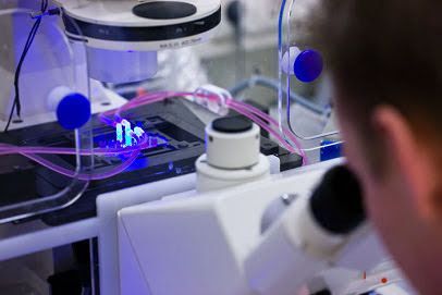
Leukocyte extravasation into inflamed tissue is a complex process that is difficult to capture as a whole in vitro. We employed a blood-vessel-on-a-chip model in which human endothelial cells were cultured in a tube-like lumen in a collagen-1 matrix. The vessels are leak tight, creating a barrier for molecules and leukocytes. Addition of inflammatory cytokine TNF-α (also known as TNF) caused vasoconstriction, actin remodelling and upregulation of ICAM-1. Introducing leukocytes into the vessels allowed real-time visualization of all different steps of the leukocyte transmigration cascade, including migration into the extracellular matrix. Individual cell tracking over time distinguished striking differences in migratory behaviour between T-cells and neutrophils. Neutrophils cross the endothelial layer more efficiently than T-cells, but, upon entering the matrix, neutrophils display high speed but low persistence, whereas T-cells migrate with low speed and rather linear migration. In conclusion, 3D imaging in real time of leukocyte extravasation in a vessel-on-a-chip enables detailed qualitative and quantitative analysis of different stages of the full leukocyte extravasation process in a single assay. This article has an associated First Person interview with the first authors of the paper.
Jaap van Buul
Transendothelial Migration
Van Buul Lab
Prof. Jaap van Buul, PhD
Biosketch
Jaap D. van Buul is Professor of Vascular Cell Biology at the University of Amsterdam, both at the faculty of Medicine and Life Sciences. He did his postdoc in the lab of Prof. Dr. Keith Burridge at the department of Developmental and Cell Biology at the University of North Carolina in Chapel Hill, USA. He showed for the first time a functional role for the small GTPase RhoG in TEM. In 2009, he co-founded the Dutch Endothelial Biology Society (DEBS), a society that gives a platform to young investigators in the vascular biology field to present their work to a larger audience and currently holds the chair. In addition, Prof. Van Buul is the president of the Dutch Society for Cell Biology (DSCB). In 2016, he joined the board of the European Vascular Biology Organization (EVBO) and in 2018 became a member of the F1000. Currently, Prof. Van Buul runs the Vascular Cell Biology lab at the Amsterdam UMC at the Department of Medical Biochemistry and is closely affiliated with the Department of Molecular Cytology at the Swammerdam Institute of Life Sciences and LCAM - van Leeuwenhoek Centre for Advanced Microscopy.
The Van Buul Lab has a long-lasting interest to understand leukocyte transendothelial migration (TEM) process and how the vessel wall manages to maintain its integrity during this event. TEM is crucial during several (patho)physiological conditions, such as inflammation, atherosclerosis, but also during homing of hematopoietic stem cells after chemo-/radiotherapy and for cancer cell metastasis.
Van Buul Lab
News
Van Buul Lab
Research
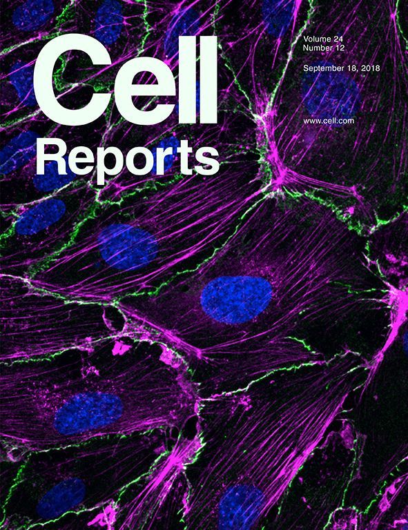
Endothelial junctional membrane protrusions serve as hotspots for neutrophil transmigration
Upon inflammation, leukocytes rapidly transmigrate across the endothelium to enter the inflamed tissue. Evidence accumulates that leukocytes use preferred exit sites, alhough it is not yet clear how these hotspots in the endothelium are defined and how they are recognized by the leukocyte. Using lattice light sheet microscopy, we discovered that leukocytes prefer endothelial membrane protrusions at cell junctions for transmigration. Phenotypically, these junctional membrane protrusions are present in an asymmetric manner, meaning that one endothelial cell shows the protrusion and the adjacent one does not. Consequently, leukocytes cross the junction by migrating underneath the protruding endothelial cell. These protrusions depend on Rac1 activity and by using a photo-activatable Rac1 probe, we could artificially generate local exit-sites for leukocytes. Overall, we have discovered a new mechanism that uses local induced junctional membrane protrusions to facilitate/steer the leukocyte escape/exit from inflamed vessel walls.

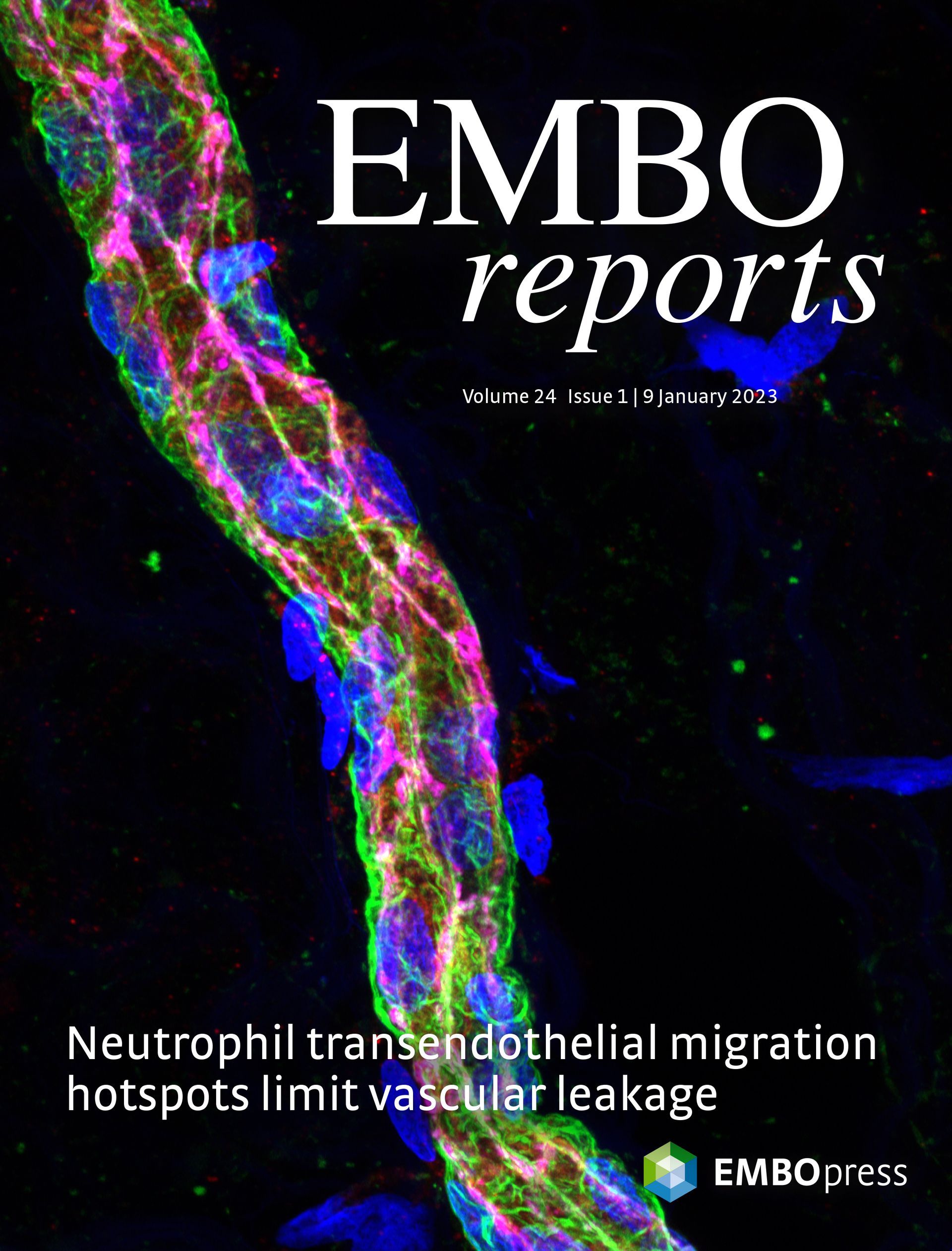
Endothelial transmigration hotspots limit vascular leakage through heterogeneous expression of ICAM-1
Upon inflammation, leukocytes leave the circulation by crossing the endothelial monolayer at specific transmigration "hotspot" regions. Although these regions support leukocyte transmigration, their functionality is not clear. We found that endothelial hotspots function to limit vascular leakage during transmigration events. Using the photoconvertible probe mEos4b, we traced back and identified original endothelial transmigration hotspots. Using this method, we show that the heterogeneous distribution of ICAM-1 determines the location of the transmigration hotspot. Interestingly, the loss of ICAM-1 heterogeneity either by CRISPR/Cas9-induced knockout of ICAM-1 or equalizing the distribution of ICAM-1 in all endothelial cells results in the loss of TEM hotspots but not necessarily in reduced TEM events. Functionally, the loss of endothelial hotspots results in increased vascular leakage during TEM. Mechanistically, we demonstrate that the 3 extracellular Ig-like domains of ICAM-1 are crucial for hotspot recognition. However, the intracellular tail of ICAM-1 and the 4th Ig-like dimerization domain are not involved, indicating that intracellular signaling or ICAM-1 dimerization is not required for hotspot recognition. Together, we discovered that hotspots function to limit vascular leakage during inflammation-induced extravasation.
Stiffness-Induced Endothelial DLC-1 Expression Forces Leukocyte Spreading through Stabilization of the ICAM-1 Adhesome
Leukocytes follow the well-defined steps of rolling, spreading, and crawling prior to diapedesis through endothelial cells (ECs). We found increased expression of DLC-1 in stiffness-associated diseases like atherosclerosis and pulmonary arterial hypertension. Depletion of DLC-1 in ECs cultured on stiff substrates drastically reduced cell stiffness and mimicked leukocyte transmigration kinetics observed for ECs cultured on soft substrates. Mechanistic studies revealed that DLC-1-depleted ECs or ECs cultured on soft substrates failed to recruit the actin-adaptor proteins filamin B, α-actinin-4, and cortactin to clustered ICAM-1, thereby preventing the ICAM-1 adhesome formation and impairing leukocyte spreading. This was rescued by overexpressing DLC-1, resulting in ICAM-1 adhesome stabilization and leukocyte spreading. Our results reveal an essential role for substrate stiffness-regulated endothelial DLC-1, independent of its GAP domain, in locally stabilizing the ICAM-1 adhesome to promote leukocyte spreading, essential for efficient leukocyte transendothelial migration.
Van Buul Lab
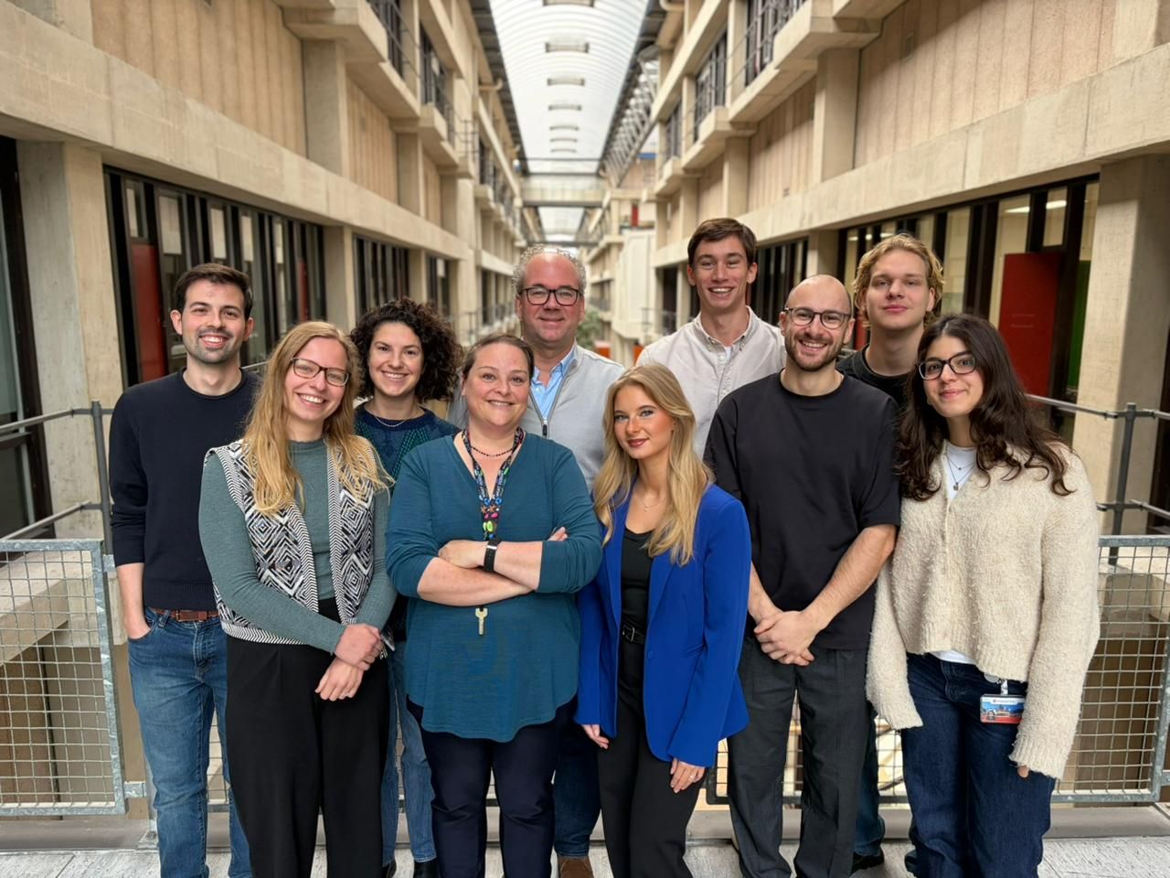
The team
Jaap van Buul
Principal Investigator

Anne-Marieke van Stalborch
Lab Manager

Nico Schramma
PostDoc
Merel Tebbens
PhD student

Werner van der Meer
PhD candidate
Anouk van der Linden
PhD student
Jeffrey van der Krogt
PhD student
Marianthi Kotsi
PhD Student

Sebastián Palacios Martinez
PhD student
Olaf Klein
Master Student
Nikki Bosmans
Master Student

Else Maas
Master Student

Gwenn Roos
Bachelor Student
Andreas Pollet
Postdoc (Tu/e)
Jos van Rijssel
Postdoc (Location Sanquin)
Van Buul Lab Alumni
* Jeffrey Kroon, ass. prof. at Amsterdam UMC, NL
* Ilse Timmerman, ass prof. at Princess Maxima Center, Utrecht, NL
* Niels Heemskerk, Research Associate at Cancer biology and immunology, Amsterdam UMC, NL
* Lilian Schimmel, Research Fellow at the Institute for Molecular Bioscience, Adelaide, Australia
* Sofia Morsing, Postdoctoral Researcher at Göteborgs Universitet, Sweden
* Anna Daniel, Medical Advice and Communications Specialist at Janssen Pharmaceuticals
* Bram van Steen, Postdoctoral Research Fellow at Wyss Institute at Harvard University
* Lanette Kempers, Postdoc at Rutgers University, Newark, USA
* Eike Mahlandt, Postdoc at NKI, Amsterdam, NL
* Janine Arts, AIOS Klinische Chemie · Diakonessenhuis, Utrecht, NL
* Max Grönloh, Postdoctoral Researcher at Utrecht Medical Center, NL
Join the Van Buul Lab
Are you interested in how leukocytes leave the vasculature and make their way through the endothelial lining, and want to work as an undergraduate or graduate student and you are highly motivated:
don't hesitate and contact us!!
Videos Van Buul Lab
Watch the videos by the Van Buul Lab.
(In Dutch)
Video in Dutch
Video in Dutch
Video in Dutch
Video in Dutch
During inflammation, leukocytes leave the circulation and cross the endothelium to fight invading pathogens in underlying tissues. This process is known as leukocyte transendothelial migration. Two routes for leukocytes to cross the endothelial monolayer have been described: the paracellular route, i.e., through the cell-cell junctions and the transcellular route, i.e., through the endothelial cell body. However, it has been technically difficult to discriminate between the para- and transcellular route. We developed a simple in vitro assay to study the distribution of endogenous VE-cadherin and PECAM-1 during neutrophil transendothelial migration under physiological flow conditions. Prior to neutrophil perfusion, endothelial cells were briefly treated with fluorescently-labeled antibodies against VE-cadherin and PECAM-1. These antibodies did not interfere with the function of both proteins, as was determined by electrical cell-substrate impedance sensing and FRAP measurements. Using this assay, we were able to follow the distribution of endogenous VE-cadherin and PECAM-1 during transendothelial migration under flow conditions and discriminate between the para- and transcellular migration routes of the leukocytes across the endothelium.
During inflammation, leukocytes leave the circulation and cross the endothelium to fight invading pathogens in underlying tissues. This process is known as leukocyte transendothelial migration. Two routes for leukocytes to cross the endothelial monolayer have been described: the paracellular route, i.e., through the cell-cell junctions and the transcellular route, i.e., through the endothelial cell body. However, it has been technically difficult to discriminate between the para- and transcellular route. We developed a simple in vitro assay to study the distribution of endogenous VE-cadherin and PECAM-1 during neutrophil transendothelial migration under physiological flow conditions. Prior to neutrophil perfusion, endothelial cells were briefly treated with fluorescently-labeled antibodies against VE-cadherin and PECAM-1. These antibodies did not interfere with the function of both proteins, as was determined by electrical cell-substrate impedance sensing and FRAP measurements. Using this assay, we were able to follow the distribution of endogenous VE-cadherin and PECAM-1 during transendothelial migration under flow conditions and discriminate between the para- and transcellular migration routes of the leukocytes across the endothelium.
Our support